PEEM station at CIRCE beamline on the flight
Magnetic (XMCD) imaging with a lateral resolution of 20 nm was demonstrated in the PEEM station at the CIRCE beamline.
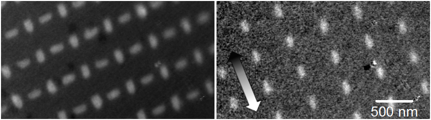
Left figure: Permalloy (NiFe) nanostructures imaged by photoelectron microscopy (PEEM) using X-rays tuned to the FeL3 absorption edge. Nanostructures of dimension 80nm*200nm*30nm (W*L*H) appear bright, while the Si background is dark (chemical contrast).
Right figure: XMCD image of the same region (difference between images with left/right handed circular polarized X-rays), showing purely magnetic contrast. The direction of contrast (parallel to incident beam) is indicated by the arrow.
Due to their shape anisotropy, all nanostructures are magnetized along their respective long axis. The XMCD image highlights magnetization along the beam direction (white), while the structures with magnetization perpendicular to the beam disappear into the background (gray). The lateral resolution (step width) in the XMCD image is 20nm, demonstrating the high resolution of the PEEM endstation at CIRCE. (Sample courtesy of P. López-Barberá & J. Nogués from ICN Barcelona and J. Perron & L. Heydermann from PSI Switzerland).
